6 Research Teams to Collaborate, Seek Cutting-Edge Discoveries
Pew Innovation Fund promotes scientific cross-pollination among biomedical program alumni
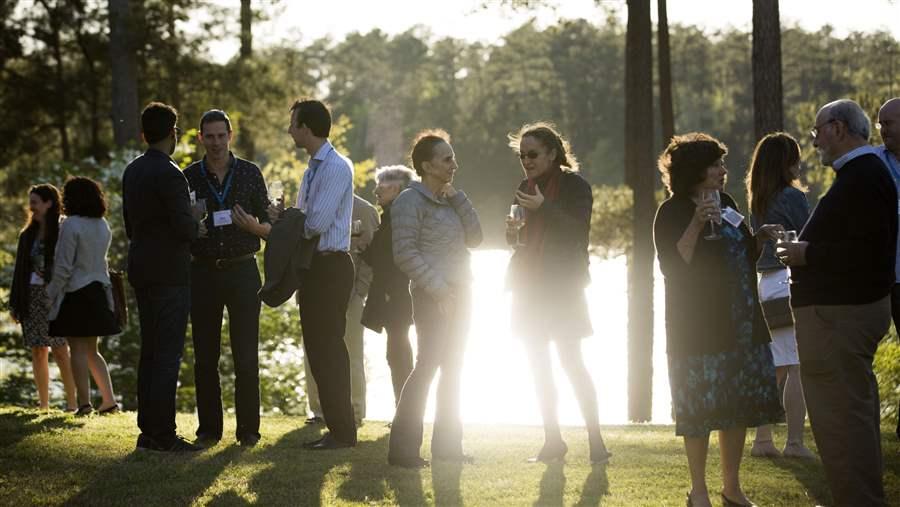
At their annual meeting, Pew’s biomedical scholars gather to connect and learn from one another. Pew’s new Innovation Fund encourages scientists from different disciplines to work together in pursuing new discoveries to benefit human health.
The Pew Charitable Trusts
Some of the most cutting-edge discoveries often occur when scientists from different backgrounds and perspectives combine their knowledge and expertise. The Pew Charitable Trusts has long encouraged collaboration among the researchers who participate in our biomedical science programs. Now, six partnerships forged by alumni of Pew’s programs will receive financial support to pursue scientific advances as the inaugural class of Innovation Fund investigators.
For over 30 years, Pew has supported more than 900 early career researchers in the United States and Latin America. The breadth of research among these scientists is unique, spanning bioengineering and neuroscience, immunology and developmental biology, and more.
The 2017 Innovation Fund awardees are building interdisciplinary research projects that combine unique areas of expertise in an effort to solve some of today’s most challenging questions in biomedical research and further drive innovation in the biomedical sciences. Their research bridges vastly different areas—from cancer biology and neuroscience to structural and developmental biology.
The 2017 Innovation Fund teams are bringing their expertise together to explore a variety of research areas, including:
How cancer cells spread
Gilad Barnea, Ph.D., and Ben Z. Stanger, M.D., Ph.D., hope to develop novel means to track cancer cells as they spread. Barnea, a neuroscientist who studies how the mammalian brain processes smell, has created a technology that maps cellular connections in the nervous system. Stanger, a developmental and cancer biologist, is keen to use the technology to track how tumors spread. The pair aims to expand the utility of this unique tracking tool to trace a cancer cell’s movement and contact with other cells—work that could help to better define how and when metastatic disease takes hold.
Intersections of neuroscience and immunology
Robert C. Froemke, Ph.D., and Dan R. Littman, M.D., Ph.D., will explore new topics at the interface between neuroscience and immunology, employing techniques from one system to investigate the other. Froemke is a neuroscientist who specializes in innovative approaches to understanding the impact of neural chemicals on animal behavior. Littman, an immunologist known for his expertise on T-cells and HIV, is interested in learning about how intestinal bacteria trigger autoimmune diseases. Together they will use novel tools to probe how the neuronal sensing of gut microbes and intestinal function can alter an animal’s behavior—work that could help map how information is relayed from the gut to the nervous system to promote recovery from an illness.
Proteins customized to study biological processes
Barbara L. Golden, Ph.D., and Andrej Luptak, Ph.D., both study RNA and through this collaboration they will combine Golden’s expertise in structural biology and catalysis with Luptak’s background in chemistry and bioengineering to develop a programmable RNA enzyme—or ribozyme—that can be used to build new customized proteins. The idea, born in Golden’s laboratory, will allow researchers to artificially incorporate specific markers and modify proteins for diverse biotechnological purposes (e.g., protein tracking and drug enhancement) and enhance the study of fundamental biological processes.
Bacteria and immune system responses
Immunologists Ivaylo I. Ivanov, Ph.D., and Pamela J. Bjorkman, Ph.D., employ vastly different approaches to investigate immune responses. Ivanov is interested in learning how the immune system responds to indigenous bacteria that occupy the gut. These bacteria play a crucial role in human health, producing essential vitamins, aiding in digestion, and preventing the growth of more harmful organisms. Bjorkman uses electron tomography—a microscopy technique for obtaining fine 3-D structures of cells—to study viral infection in tissues. While seated together at an annual Pew meeting, the two discussed ways to bring together their complementary expertise, which sparked the idea of performing structural characterizations of the interactions between noninvasive microbes and intestinal cells. Findings from this research will allow Bjorkman and Ivanov to learn how bacteria interact with the intestine to modulate immune responses, and identify mechanisms that help regulate normal bodily functions.
How viruses replicate and persist
Benjamin R. tenOever, Ph.D., and Felicia D. Goodrum, Ph.D., both 2008 alumni, have a common interest in viruses and is looking for ways to better understand how viruses cause infections. TenOever’s research is focused on using RNAs to control virus infections and develop novel therapeutic tools. Goodrum is dedicated to understanding how the herpesvirus can stay in the host to cause lifelong latent infection. This grant allows them to combine efforts in developing a system to study human cytomegalovirus (HCMV)—a form of herpesvirus—in cell types important to viral persistence. Goodrum and tenOever will devise a system to better manipulate the virus to tease out the function of specific viral genes. Their findings are poised to advance understanding of how viruses lie dormant to cause persistent infections and may ultimately result in new ways of controlling virus replication and developing an effective HCMV vaccine.
3-D views of cell pathways
William I. Weis, Ph.D., and Georgios Skiniotis, Ph.D., bring a unique set of skills to study Wnt signaling—a pathway implicated in cellular development and cancer. Weis’ lab is immersed in Wnt research and uses biochemistry and biophysics-based tools to study the structure of Wnt signaling components, while the Skiniotis lab specializes in cryo-electron microscopy—a microscopy technique used to observe samples at a very low temperature—to dissect the structure of proteins. Together, Weis and Skiniotis will perform studies to obtain a 3-D structure of a major Wnt complex where various pathway components come together to form an organized scaffold. This is of particular interest because this complex is a major hub of Wnt activity, and understanding its assembly could lead to insights on how the pathway is regulated.
Kara Coleman directs The Pew Charitable Trusts’ biomedical programs, including the biomedical scholars, Pew-Stewart Scholars for Cancer Research, and Latin American fellows programs.


